Friday, January 31, 2014

Yersinia, Francisella, Brucella and Pasteurella (Gram-Negative)

These are gram-negative rods that cause disease in animals, and are transferred from animals to humans. They are facultative intracellular organisms (with the exception of Pasteurella multocida), meaning the bacteria can survive within macrophages, set up infection to regional lymph nodes of the site of contact, and infect organs via the bloodstream. Immune response is cell-mediated and exhibits delayed-type hypersensitivity.

Yersinia pestis
Causes bubonic plague. (Think rat and "pest"). This disease is still contractible in southwestern US. 
  • Virulence factors: 
    • Fraction 1 (F1), antiphagocytic capsular antigen.
    • V and W antigens, unknown function.

Francisella tularensis 
Causes tularemia, which closely resembles the bubonic plague. Infects many mammals, especially rabbits, and is carried by ticks and deerflies.
  • Diseases: 
    • Tularemia. Like the bubonic plague, symptoms include fever, swollen lymph nodes, and infection of multiple organs. Unlike the bubonic plague, there is no skin ulcer. 

Brucella
Usually contracted from handling meat, and is common worldwide in countries where animals are not immunized or where milk is not pasteurized.

Pasteurella multocida
Not a facultative intracellular organism. Lives in the mouths of cats, and infects humans from cat or dog bite.

Thursday, January 30, 2014

Haemophilus, Bordetella and Legionella (Gram-Negative)

These bacteria infect the respiratory tract.

Haemophilus influenzae
Grows in the blood. Infects the lungs of patients who are weakened by the viral flu infection, and infects children who are at the stage of developing their immune system.
  • Virulence factors:
    • Polysaccharide capsule, of which there are six types: a, b, c, d, e and f. Capsule b is associated with the disease in children. Bacteria lacking a capsule can only cause local infection.
  • Child diseases (type b):

Haemophilus ducreyi
Causes the STD, chancroid.

Gardnerella vaginalis
Causes bacterial vaginitis with the presence of anaerobic vaginal bacteria.

Bordetella pertussis
Causes disease by toxin release, not bacteria-cell invasion.
  • Exotoxins:
    • Pertussis toxin - like most exotoxins, its B subunit binds to the target cell receptors and gain entry. The A subunit activates G protein in target cell membrane and, in turn, activates intracellular messengers such as cAMP. In the case of pertussis, this signal causes:
      • Histamine sensitization
      • Increased insulin synthesis
      • Increased lymphocyte production and inhibition of phagocytosis
    • Extra cytoplasmic adenylate cyclase - this toxin is taken up by host neutrophils, lymphocytes and monocytes. Adenylate cyclase synthesizes cAMP, and in this case, weakens the defense cells' ability to lyse and phagocytose.
    • Filamentous hemagglutinin (FHA) - pili that are responsible for attachment to bronchi epithelium.
    • Tracheal cytotoxin - destroys ciliated epithelial cells, causing more buildup of mucus and bacteria.
  • Diseases:

Legionella pneumophila
Infection by airborne bacteria from contaminated water, but not person-to-person transmission. It survives phagocytosis and replicates intracellularly.

Wednesday, January 29, 2014

Hospital-acquired Gram-Negative Bacteria

Pseudomonas aeruginosa
The most problematic. Only infects weakened humans, and is very resistant.
  • Diseases (BEPSEUDO):
    • Burn-wound infections.
    • Endocarditis.
    • Pneumonia by air contamination.
    • Sepsis from infected catheters or from other sites of infection. 
    • External malignant otitis from an external ear canal infection. 
    • Urinary tract infection. 
    • (Diabetic) Osteomyelitis by wound infection, especially in diabetics who develop foot ulcers. 
    • Corneal infections in contact lens wearers. 

Burkholderia cepacia
Transmitted to patients from water and drug resistant.

Stenotrophomonas maltophilia
Aerobic, part of normal respiratory flora, but can cause disease in immunocompromised patient.

Acinetobacter
Aerobic, causes a wide range of infections.

Tuesday, January 28, 2014

Enterics (Gram-Negative)

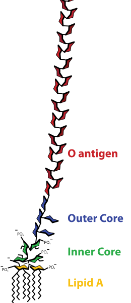
These are bacteria that live in the intestine, but can cause infection and disease. The enterics have varying but common cell surfaces: O antigen of the LPS, covered by K antigen capsule. H antigen, which is a subunit of the flagella, is present only in motile bacteria.

The main family groups are enterobacteriaceae, vibrionaceae, pseudomonadaceae and bacteroidaceae. Some groups contain many species, and are simply mentioned by their genus.

Pathogenesis
  • Causes of diarrhea:
    1. Release of exotoxins without entering cells of the GI tract. The intestinal epithelium reacts by losing electrolyte and fluid, causing watery diarrhea without symptoms. 
    2. Invasion of intestinal epithelium destroys the cells by toxins and causes bleeding. Immune response causes fever. 
    3. Invasion of the lymph and bloodstream causes abdominal pain, intestinal bleeding, fever and headache. A deeper invasion can cause lymph node enlargement, bacteremia and sepsis.
  • Hospital-acquired infection - urinary tract infections, pneumonia, bacteremia and sepsis.

ENTEROBACTERIACEAE

Escheria coli
Constantly uptakes new DNA from conjugation, lysogenic conversion by bacteriophages and direct transposon mediated DNA insertion. If E. coli acquires virulence, it can cause disease, although it is part of normal flora in the intestine.
  • Diseases:
    • Diarrhea by strains: 
      • ETEC (Enterotoxigenic E. coli), cause 1 (see Pathogenesis above). Release of exotoxins heat labile toxin (LT) and heat stable toxin (ST) causes severe watery diarrhea, as by cholera.
      • EHEC (Enterohemorrhagic E. coli), cause 1. Secretes powerful Shiga-like verotoxin. Blood in diarrhea.
      • EIEC (Enteroinvasive E. coli), cause 2. Blood and pus in diarrhea, as by shigellosis.
    • Urinary tract infection - acquisition of a pili virulence factor allows bacteria to travel up the urethra and infect the bladder (cystitis) and sometimes even the kidney (pyelonephritis).
    • Neonatal meningitis.
    • Gram-negative sepsis - usual and most common cause of hospital sepsis.

Klebsiella pneumoniae
Encapsulated (O) but non-motile (no H antigen). It is present in hospitals, causing sepsis and urinary tract infections via Foley catheters. Results in violent symptoms that can destroy lung tissues.

Proteus mirabilis
Very motile. See hospital-acquired infections.

Enterobacter
Very motile. Occasionally responsible for hospital-acquired infections.

Serratia
Can cause urinary tract infections, wound infections and pneumonia. Characterized by bright red pigment.

Shigella
This group has four non-motile species (dysenteriae, flexneri, boydii and sonnei) that are actually not part of normal intestinal flora, and are always pathogenic. Bacteria invade intestinal epithelial cells, and release the Shiga toxin, which disables cells' ability to reabsorb fluids and electrolytes. Diarrhea cause 2 (see Pathogenesis above).

Salmonella
Motile. Has Vi antigen capsule, similar to the K antigen, which surrounds the O antigen to protect it against antibody attack. Not part of normal intestinal flora, and always pathogenic. Lives in GI tracts of animals, and only infects humans when there is contamination of food or water with animal feces.
  • Diseases (big 4):
    • Typhoid fever.
    • Carrier state - some people recovering from typhoid fever can become chronic carriers. They keep Salmonella typhi in their gallbladders, excrete the bacteria constantly and display no symptoms. Recall Typhoid Mary. 
    • Sepsis - bacteria in bloodstream carried to lungs, brain or bone. This is usually an infection of Salmonella choleraesuis and does not involve GI tract.
    • Gastroenteritis (diarrhea) - most common symptom of this bacterium, caused by a cholera-like toxin that causes watery diarrhea (cause 1) with trace mucous and blood. 

Yersinia enterocolitica
Not an enteric bacterium, but causes diarrhea (cause 3). Ingested from contaminated foods, such as milk for fecally contaminated water. Secretes exnterotoxin, similar to heat-stable senterotoxin of e coli. Fever, diarrhea, abdominal pain.


VIBRIONACEAE

Vibrio cholera
Causes cholera, a diarrheal disease (cause 1) from infection by fecal contamination. Multiplies in intestine, causing similar but more severe form of ETEC (see Escheria coli above). Toxin is called choleragen, with the same mechanism as LT toxin.

Vibrio parahaemolyticus
A marine bacterium that causes gastroenteritis after ingestion of uncooked seafood (sushi).

Campylobacter jejuni
One of the most common causes of diarrhea in the world, along with ETEC and rotavirus. Found in animals and infect humans by contamination of food or water.

Helicobacter pylori
Most common cause of duodenal ulcers and chronic gastritis (inflamed stomach) and second-most common cause of gastric ulcers.


BACTEROIDACEAE

Bacteroides fragilis 
One of the few gram-negative bacterium that do not posses the endotoxin lipid A. This is a normally peaceful bacterium until it infects a wound and form an abscess. Fever and sometimes systemic spread accompany the infection.

Bacteroides melaninogenicus 
Lives in mouth, vagina and intestine. Causes periodontal disease and aspiration pneumonia.

Fusobacterium
Causes the same diseases as B. melaninogenicus, abdominal and pelvic abscesses and middle ear infection.

Monday, January 27, 2014

Culprits of Meningitis in Babies

Meningitis caused by infection from the birth canal:
Listeria monocytogenes
Escherichia coli ("E. coli")
Streptococcus agalactiae (Group B streptococcus)

Meningitis caused by community infection:
Neisseria meningitis
Haemophilus influenzae

Neisseria (Gram-Negative)

Neisseria are diplococci, meaning they appear as round shaped pairs. Neisseria meningitidis causes meningitis, and Neisseria gonorrhoeae causes gonorrhea.

Neisseria meningitidis
These bacteria are transmitted by respiratory secretions (saliva), and can blend into and become part of normal flora of the nasopharynx in 5-15% of adults. They affect infants of 6 months to 2 years due to their weak immune systems, or army recruits and college freshmen who are exposed to a close group environment. 
  • Virulence factors:
    • Polysaccharide capsule that is antiphagocytic. 
    • LPS endotoxin that can destroy blood vessel and causes sepsis if released. The blood vessel hemmorhage causes petechial rash, which is a classic sign of meningococcal infection.
    • IgA1 protease, an enzyme that cleaves IgA antibodies. 
    • Pili that attach to cells of the nasopharynx.
  • Diseases: 
    • Meningococcemia - sudden fevers, chills, joint and muscle pains due to multiplication of bacteria in the bloodstream.
    • Fulminant meningcoccemia - septic shock with adrenal gland hemorrhage, causing abrupt hypotension, rapid heart rate (tachycardia), worsened petechiae, disseminated intravascular coagulation (DIC) and death. 
    • Neonatal meningitis (late-onset) - most common form of meningococcal disease. Causes fever, vomiting and irritability.

Neisseria gonorrhoeae 
The second most common cause of STD. Transmitted as long as there is a transmission of body fluids: sexually, orally or anally.
  • Virulence factors:
    • Pili that are hypervariable as a protection against antibodies or vaccines. Pili adhere to host cells so strongly that phagocytosis can be prevented.
    • Outer membrane protein porins promote invasion into epithelial cells.
    • Opa proteins of the outer membrane promote adherence and invasion into epithelial cells. 
    • LPS can destroy cilia of cells before the bacterium is taken up by endocytosis. 
  • Diseases: 
    • Gonococcal bacteremia when a rare invasion of the bloodstream occurs. Fever, joint pains, skin lesions, followed by rare occurrence of heart inflammation (endocarditis and pericarditis) or meningitis after the bacteria spreads.
    • Septic arthritis causing acute fever and pain of one or two joints.
    • In men: the urethra is the site of infection. Inflammation of the urethra (urethritis) causes painful urination, pus discharge, and possibly epididymitis, prostatitis and urethral strictures (narrowing of the urethra).
    • In women: a less symptomatic version of urethritis in males, such as lower abdominal pain or no symptoms at all. Infection of the cervix epithelium can progress to pelvic inflammatory disease (PID).
    • In infants: infection during delivery, resulting in eye infection (ophthalmia neonatorum) and possible blindness.

Saturday, January 25, 2014

Bacillus, Clostridium, Corynebacterium and Listeria (Gram-Positive)

This entry is on the medically important gram-positive bacteria that are rods. They are categorized as spore-forming (bacillus and clostridium) and non-spore-forming (corynebacterium and listeria).

Bacillus anthracis
The only bacterium with a capsule made of protein called poly-D-glutamic acid. Commonly known as anthrax, which infects cows and sheep and is then transferred to humans by contact. Its spores are very hardy and germinate inside the body, releasing exotoxins. Diseases can be treated with penicillin.
  • Diseases:
    • Cutaneous anthrax from spore germination on skin wounds. 
      • Exotoxin release causes localized tissue necrosis that can progress to skin edema and shock in ~10% of cases. Spore proliferation in the bloodstream can cause death. 
    • Pulmonary aka Respiratory anthrax. 
      • Macrophages take up spores in the lungs, and transport them to hilar and mediastinal lymph nodes at the bronchi and heart, respectively. There, spores proliferate and cause mediastinal hemorrhage and pleural effusions.
    • GI anthrax
      • Ingestion of spores cause necrotic lesion in the intestine. Results in vomiting, abdominal pain and bloody diarrhea.
  • Exotoxin proteins:
    • Edema factor (EF) increases cAMP, which impairs neutrophil function and disrupts water homeostasis (edema).
    • Protective antigen (PA) promotes entry of EF into phagocytic cells.
    • Lethal factor (LF), enzyme that inactivates protein kinase, and stimulates macrophages to release TNF and IL-1β.

Bacillus cereus
Causes food poisoning due to the bacteria depositing spores and enterotoxin (an exotoxin that targets intestine) into food. Bacteria are not encapsulated, and are resistant to penicillin.

Clostridium botulinum
Produces a lethal neurotoxin that causes rapid and severe food poisoning. The neurotoxin blocks ACh release, leading to muscle paralysis.
  • Diseases:
    • Adult botulism, with difficulty swallowing (dysphogia), double vision caused by bilateral cerebral nerve palsies, and muscle weakness leading to paralysis and death. Respiratory failure from poor ventilation.
    • Infant botulism, with initial constipation then difficulty swallowing and muscle weakness. Prognosis is better than that of an adult.

Clostridium tetani
Commonly found in soil and animal feces, and causes tetanus. Spores deposited in wounds germinate in an anaerobic environment, such as necrotic tissue. The release of exotoxin tetanospasmin, taken up by the neuromuscular junction to the CNS, prevents the release of inhibitory neurotransmitters GABA and glycine. This results in a high frequency of impulses and a sustained contraction. Diseases include respiratory failure, lockjaw and other severe muscle spasms.

Clostridium perfringens
Anaerobic, found in soil, releases exotoxin enzymes and produces gas.
  • Diseases:
    • Cellulitis/wound infection, where bacterial growth from necrotic skin damages local tissue.
    • Clostridial myonecrosis in the muscle, damage by exotoxins and fatal unless treated early. Other enzymes released by the bacteria form gas bubbles within muscle and subcutaneous tissue.

Clostridium difficile
After the use of broad spectrum antibiotics, which can wipe out intestinal flora, the colonization of the C. difficile and the release of its exotoxins cause severe diarrhea, cramps and fever.

Corynebacterium diphtheriae
Colonizes the pharynx, where the bacteria forms a pseudomembrane of fibrin, leukocytes, necrotic epithelial cells and C. diptheriae cells. Exotoxin release at this site into the bloodstream interferes with protein synthesis in the heart and CNS. Causes diphtheria.

Listeria monocytogenes
An exception to the pattern of gram-positive bacteria, L. monocytogenes possesses endotoxin. They can hide within macrophages and neutrophils, and are likely to invade infants and immunosuppressed patients whose immune systems are weak. They are one of the common causes of meningitis.

Thursday, January 23, 2014

Staphylococci (Gram-Positive)

Staphylococci appear as clusters (think "staff"). Most are penicillin G resistant. Three major pathogenic species are Staphylococcus aureus, Staphylococcus epidermidis and Staphylococcus saprophyticus.

Methods of differentiation from streptococci
  1. Gram staining to reveal physical traits. 
  2. Catalase test to show enzyme catalase, which is present in all staphylococci. 
  3. Culture to show hemolysis ability. Most staphylococci are either beta-hemolytic or not hemolytic. Staphylococcus aureus leaves a gold pigment on sheep blood agar. Remember alpha-hemolytic bacteria leave a green pigment on blood agar.

Of the pathogenic species, only S. aureus is coagulase-positive
Most S. aureus produces the enzyme coagulase, which clots the blood. Fibrin formation around the bacterium can protect it from phagocytosis.

Staphylococcus aureus
  • Defensive proteins: 
    • Protein A binds IgG at the Fc region (the tail rather than the arms), preventing opsonization. 
    • Coagulase.
    • Hemolysins alpha, beta, gamma and delta, which destroy RBCs, neutrophils, macrophages and platelets.
    • Leukocidins destroys leukocytes. 
    • Penicillinase inactivates penicillin. 
    • Novel penicillin binding protein (transpeptidase) forms the cell wall, and is inhibited by penicillin. 
  •  Tissue "tunneling" proteins:
    • Hyaluronidase ("Spreading Factor") breaks down proteoglycans in connective tissue. 
    • Staphylokinase breaks down fibrin clots. 
    • Lipase degrades fats and oils, allowing colonization in sebaceous glands. 
    • Protease destroys tissue proteins. 
  • Exotoxins:
    • Exfoliatin causes the detachment in the epidermal layer, leaves blisters.
    • Enterotoxin causes food poisoning, vomiting and diarrhea. 
    • Toxic Shock Syndrome toxin is a superantigen that binds to MHC II of APCs, causing a massive T cell response (comparable to but deadlier than pyrogenic toxin). 
  • Diseases:

Staphylococcus epidermidis 
Part of normal bacterial flora and lives widely on the skin without causing disease. However, it causes infections if it enters wounds on medical devices such as artificial hip joints or heart valves, or in the bloodstream via catheters.

Staphylococcus saprophyticus 
Causes urinary tract infection in sexually active young women. 

Tuesday, January 21, 2014

Streptococci (Gram-Positive)

Alpha and Beta haemolytic streptococci by GrahamColm
Streptococci, coccus meaning "sphere", appear as strips of balls. Unlike staphylococci, streptococci does not have enzyme catalase.

Classification
By the ability to lyse red blood cells: Beta, completely hemolytic. Alpha, partially hemolytic and leaves a greenish color on the culture medium around the colony. Gamma, not hemolytic.

By Lancefield antigens found in the cell wall, which differentiate species: Group A (beta), B (beta) and D (alpha). Include Streptococcus pneumoniae (alpha) and the Viridans group (alpha), which make up the five pathogenic streptococci species in humans.


Group A Streptococcus pyogenes
  • Beta-hemolytic.
  • Cell wall antigens:
    • M protein prevents complement activation, but can be targeted by antibodies.
  • Pathogenic enzymes:
    • Streptolysin O destroys RBCs and white blood cells. Oxygen labile (inactivated by oxygen).
    • Streptolysin S destroys RBCs and white blood cells. Oxygen stabile.
    • Pyrogenic exotoxin are "super" antigenic, causes scarlet fever and toxic shock. Only present in a few strains.
    • Streptokinase activates plasmin, an enzyme that breaks up blood clots.
  • Diseases: 

Group B Streptococcus agalactiae
  • Beta-hemolytic. 
  • Diseases (of babies):
    • Meningitis, pneumonia, sepsis by infection from the vagina after birth.

Group D Enterococcus faecalis, Enterococcus faecium, Non-enterococci (Streptococcus bovis, Streptococcus equinus, ...)
  • Alpha-hemolytic. 
  • Strains resist most drugs, live in the GI tract, and cause infections of the urinary tract or biliary tract. 

Streptococcus pneumoniae
  • Alpha-hemolytic. 
  • Antigens:
    • Polysaccharide capsule prevents bacterium from being phagocytized.
  •  Diseases:

Viridans Streptococcus salivarius, Streptococcus sanguis, Streptococcus mitis, ...
  • Alpha-hemolytic.
  • Diseases:
    • Dental infections and cavities.
    • Endocarditis by dental infection leading to bacterial growth in the heart valves via bloodstream.
    • Abscesses in the abdomen or brain, caused by Streptococcus intermedius.

Monday, January 20, 2014

Gram-Positive and Gram-Negative Bacteria

Based on their cell wall structure, bacteria are either gram-positive or gram-negative as result of the gram stain (animation). "Negative" here meaning the initial crystal violet dye has not been retained so the lighter colored counter stain is more apparent.

Comparisons, Layers of the cell envelope, in order: (Image search)
Gram-Positive
  1. Extracellular.
  2. Thick cell wall, allowing more cross-link of amino acid side chains. 
    • Presence of teichoic acid - a polysaccharide that can be used to identify gram-positive bacteria.
  3. Plasma membrane. 
    • No endotoxin or porin channel in plasma membrane.  
  4. Intracellular.

Gram-Negative 
  1. Extracellular. 
  2. Outer plasma membrane. 
    • Porin channels help the passage of nutrients. 
    • Outermost lipid bilayer has lipopolysaccharide (LPS) instead of phospholipid.
    • LPS contains an endotoxin called lipid A. Lysed gram-negative cells release lipid A into circulation, causing fever, diarrhea and septic shock.
  3. Periplasmic space / Murein lipoprotein, which binds the outer membrane to the cell wall layer.
  4. Thin cell wall / Periplasmic space. 
    • No teichoic acid in cell wall. 
  5. Inner plasma membrane.
  6. Intracellular.

A thought on gram staining
Crystal violet and iodine are small enough to pass through cell membrane or cell wall. The subsequent complexes they make in the cell envelope is large, however still passable through porins. Alcohol strips away the outer membrane of gram-negative cells, allowing the complexes to leave past the lipid membrane.
LPS by Mike Jones

Gram-negative species are more dangerous
The outer LPS-containing membrane blocks antibiotics and chemicals from attacking the cell wall. This makes the bacterium resistant to lysosome and penicillin. The presence of endotoxin makes gram-negative bacteria more pathogenic than gram-positive bacteria.

Lipopolysaccharides (LPS)
0-antigen is a side chain specific to different organisms - think O for Outer. Lipid A is a disaccharide with fatty acid tails.

Transpeptidase 
An enzyme that cross-links the bacterial cell wall side chains.