 |
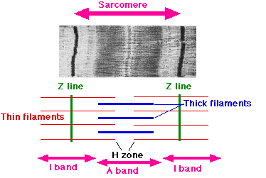
| Sarcomere by Sameerb |
Mechanoreceptors
Golgi tendon organs detect tension in muscle tendons. Muscle spindles lie within the center of a muscle, and detect stretch.
These receptors provide continuous feedback on the degree of muscle stretch and tension and the speed of stretching in order to achieve smooth and precise muscle action.
Muscle spindle
The two sensory receptors of muscle spindles are called primary and secondary nerve endings. They relay information about the degree of muscle stretching. Primary nerve endings also detect the rate of muscle stretching.
If a muscle remains stretched, the receptors send negative feedback information to the spinal cord and cerebellum, causing muscle contraction. If a muscle is suddenly stretched, then the primary receptors provide additional strong input to the spinal cord. An example is the knee jerk reflex.
Muscle spindles also consist of extrafusal muscle fibers (stimulated by gamma motor neurons), and smaller intrafusal muscle fibers (alpha motor neurons). Gamma motor neurons fire whenever alpha motor neurons fire, ensuring the length of intrafusal fibers and extrafusal fibers keep pace in contraction.
Golgi tendon organ
The Golgi tendon organ reacts against increased muscle tension by decreasing tension (allowing muscle to stretch).
Muscle contraction (animation)
- An action potential spreads through the muscle fiber's T-tubules network.
- Depolarization of the muscle fiber eventually causes the sarcoplasmic reticulum to release calcium.
- The calcium binds to the actin filament (on troponin C), causing an allosteric change of troponin and allowing tropomyosin to move. The binding site is unblocked. When calcium is present the blocked active site of the actin clears.
- Myosin binds to the binding site on the thin filament, releasing phosphate and then ADP. (The myosin head has ADP and Pi attached in its high energy configuration).
- The release of ADP is coupled to the power stroke, where the myosin head pivots and pulls the actin filament toward the center.
- ATP binds to myosin head, allowing it to release actin and be in the weak binding state. The lack of ATP results in the rigor state characteristic of rigor mortis.
- ATP is split into ADP and Pi by hydrolysis.
- Steps 4 to 7 repeat as long as ATP is available and calcium is freely found within the thin filaments.
- Meanwhile, calcium is actively pumped back into the sarcoplasmic reticulum. When calcium is no longer present on the thin filament, the tropomyosin changes conformation back to its previous state, and blocks the binding site again. The myosin ceases binding to the thin filament, and the contractions cease.
No comments:
Post a Comment